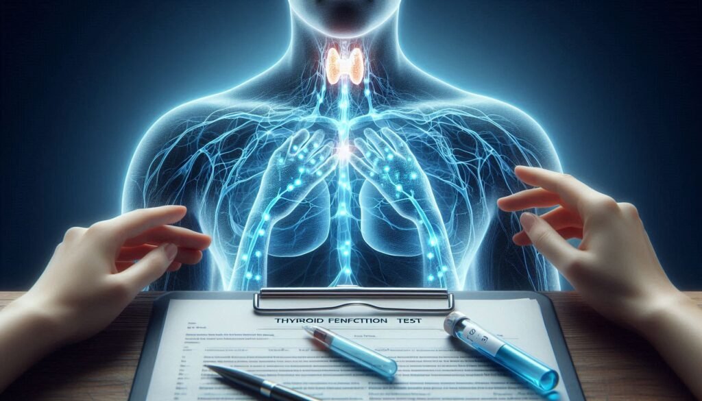
Paresthesia can be a perplexing sensation. It often manifests as tingling, prickling, or numbness in various parts of the body. While it might seem harmless at first, this symptom can signal underlying neurological issues that require careful evaluation. Understanding what’s happening beneath the surface is crucial for effective treatment and management.
Neurological exams play a vital role in diagnosing paresthesia. They help healthcare professionals identify potential nerve dysfunction and pinpoint its causes. Whether it’s related to injuries, diabetes, multiple sclerosis, or other conditions, these assessments provide valuable insights into your nervous system’s health.
In this article, we’ll explore the step-by-step process of neurological exams specifically tailored for paresthesia diagnosis. From cranial nerve assessments to cognitive evaluations—each component reveals critical information about your body’s neural pathways and how they’re functioning. Let’s dive deeper into this essential aspect of medical evaluation and shed light on what you can expect during your examination journey!

The Importance of Neurological Exams in Paresthesia Diagnosis
Neurological exams are essential for accurately diagnosing paresthesia. These examinations provide a comprehensive assessment of the nervous system, helping to identify potential issues that could be causing unusual sensations. By understanding the underlying causes, healthcare providers can develop appropriate treatment plans.
Paresthesia can arise from various factors, including nerve compression, injury, or systemic diseases like diabetes and multiple sclerosis. A thorough neurological exam helps differentiate between these possibilities. This is crucial in determining whether paresthesia is a symptom of something benign or indicative of a more serious condition.
During the exam, clinicians assess function at different levels—ranging from cranial nerves to motor skills and reflexes. Each component contributes valuable information about how well your nervous system operates as a whole.
Timely diagnosis through neurological exams not only aids in effective management but also alleviates patient anxiety by providing clarity on their symptoms. Understanding what’s happening neurologically empowers individuals to make informed decisions about their health journey.
Cranial Nerve Assessment: Testing Facial Sensations
Cranial nerve assessment plays a critical role in understanding paresthesia, especially when it affects facial sensations. The face is innervated by several cranial nerves, notably the trigeminal nerve (cranial nerve V), which is responsible for sensory input to different areas of the face. Testing this nerve helps identify any abnormalities that could indicate underlying neurological issues.
During an examination, clinicians often use light touch and pinprick tests to evaluate facial sensation across three distinct branches: ophthalmic, maxillary, and mandibular. Patients are asked to close their eyes while the examiner lightly touches various points on their face. This process helps pinpoint any regions where sensation may be diminished or altered.
Responses from patients can reveal valuable information about potential dysfunctions within these nerves. Additionally, assessing for asymmetry in sensation between sides of the face can help narrow down possible causes of paresthesia.
Furthermore, other cranial nerves involved in facial movement—such as the facial nerve (cranial nerve VII)—may also be examined concurrently to assess overall function and coordination within this region.
Muscle Strength Evaluation: Pinpointing Weakness
Muscle strength evaluation is a crucial component of neurological exams for paresthesia. It helps identify areas where weakness may be present, offering insights into underlying nerve or muscle issues. During this assessment, healthcare professionals typically ask patients to perform specific movements against resistance.
Healthcare providers often utilize manual muscle testing to gauge the strength of individual muscle groups. This involves asking patients to push or pull against their examiner’s hands while maintaining control and stability. The examiner observes both the force exerted and any compensatory movements that may indicate weakness.
Another method used is dynamometry, which quantitatively measures grip strength and other muscular functions. These measurements provide precise data on how well muscles can perform tasks under stress.
A thorough evaluation not only highlights weak areas but also assists in differentiating between peripheral nerve injuries and central nervous system disorders. Understanding these distinctions can guide further diagnostic steps and treatment options tailored for individuals experiencing paresthesia symptoms.
Reflex Testing: Assessing Nerve Responsiveness
Reflex testing is a crucial component of neurological exams for paresthesia. It evaluates how effectively the nervous system responds to stimuli. By checking reflexes, healthcare providers can identify potential issues with nerve pathways.
During this assessment, a physician typically uses a small hammer to tap specific tendons in the body. Common sites include the knee and ankle joints. The response should be quick and automatic, indicating healthy nerve function.
An exaggerated or diminished response may suggest underlying problems. For example, hyperactive reflexes could point toward central nervous system disorders, while reduced responses might indicate peripheral neuropathy or spinal cord injuries.
Additionally, reflex tests help differentiate between different types of nerve damage—sensory versus motor—as well as localize potential lesions within the nervous system. This information is vital for developing an accurate diagnosis and tailored treatment plan for those experiencing paresthesia.
Sensory Examination: Mapping Areas of Altered Sensation
A sensory examination is crucial for diagnosing paresthesia. This process involves assessing how well the nerves transmit sensations like touch, pain, and temperature. By using various tools and techniques, healthcare professionals can pinpoint specific areas where sensation may be altered.
During the assessment, doctors often use a cotton swab or a tuning fork to evaluate different types of sensory perception. They might ask patients to close their eyes while they gently touch various parts of the body. This helps identify regions with reduced sensitivity or abnormal sensations.
Mapping these areas provides valuable insights into potential nerve damage or dysfunction. For instance, if a patient reports tingling in a particular limb, identifying exactly where this sensation begins and ends can help narrow down possible causes.
Additionally, responses to stimuli are noted carefully. The presence of hyperesthesia (increased sensitivity) or hypoesthesia (decreased sensitivity) aids in creating an accurate picture of the underlying neurological issues affecting the patient’s condition.
Coordination Tests: Identifying Balance and Movement Issues
Coordination tests are crucial in evaluating how well the nervous system controls movement and balance. These assessments help identify any abnormalities that may contribute to paresthesia, or abnormal sensations in the body.
During these tests, healthcare professionals observe a patient’s ability to perform tasks requiring fine motor skills and overall coordination. Simple activities like finger-to-nose testing can reveal issues with proprioception—the body’s sense of its position in space.
Other exercises include heel-to-shin movements, which assess lower limb coordination. Abnormalities during these assessments can indicate disturbances within specific brain regions or peripheral nerves.
Balance is also evaluated through various standing and walking tasks, such as tandem walking or Romberg test. Difficulties with these movements might signal underlying neurological conditions affecting muscle control and stability, providing essential insights for accurate diagnosis.
Gait Analysis: Observing Walking Patterns
Gait analysis is a crucial component in neurological exams for paresthesia. It involves observing how a person walks to identify any abnormalities that may indicate underlying nerve issues. Noticing changes in walking patterns can provide valuable insights into the nervous system’s function.
During this assessment, healthcare professionals look for various factors such as stride length, balance, and overall coordination. An irregular gait could suggest muscle weakness or sensory deficits affecting movement. Observing these aspects helps pinpoint potential areas of concern related to nerve damage or dysfunction.
Different types of gaits are analyzed, including limping or shuffling. Each variation might point toward specific conditions impacting the nerves and muscles. The examiner often uses video recording technology to capture subtle differences that might be missed during direct observation.
Gait analysis serves as an essential diagnostic tool within neurological exams for paresthesia. It allows clinicians to formulate tailored treatment plans based on individual needs and symptoms observed during the walking evaluation process.
Autonomic Function Tests: Evaluating Involuntary Nervous System
Autonomic function tests are essential tools in assessing the involuntary nervous system. This system controls bodily functions that we often take for granted, such as heart rate, blood pressure, and digestion. For individuals experiencing paresthesia, understanding how well this system is functioning can provide insights into underlying health issues.
One common test involves measuring changes in heart rate and blood pressure while a patient shifts from lying down to standing up. This tilt table test helps identify conditions like orthostatic hypotension, which can lead to dizziness or fainting.
Another method includes sweat testing to evaluate nerve responses related to temperature regulation. Abnormal sweating patterns may indicate disruptions within the autonomic pathways.
Additionally, deep breathing exercises during assessment can be revealing. By evaluating respiratory heart rate variability, healthcare professionals gain further insight into autonomic balance and potential dysfunctions associated with paresthesia symptoms.
Cognitive Assessment: Ruling Out Neurological Disorders
Cognitive assessments are vital when evaluating patients with paresthesia. They focus on mental processes such as memory, attention, and problem-solving abilities. Neurological disorders can often manifest through cognitive impairments, making these tests crucial for accurate diagnosis.
During the assessment, healthcare professionals may use standardized tools like the Mini-Mental State Examination (MMSE) or Montreal Cognitive Assessment (MoCA). These instruments help gauge a person’s cognitive function in various areas, allowing practitioners to identify any deficits that could indicate an underlying issue.
Patients might be asked to recall words or follow multi-step instructions. Such tasks provide insight into their short-term memory and executive functioning skills. Observing how well individuals perform these activities can reveal significant information about their neurological health.
Recognizing cognitive dysfunction is essential for developing a comprehensive treatment plan. It helps differentiate between peripheral issues causing paresthesia versus central nervous system complications requiring further evaluation.
Interpreting Neurological Exam Results: What They Mean for You
Interpreting the results of neurological exams for paresthesia is crucial for understanding your condition and determining the next steps in treatment. The findings from each component of the exam can provide valuable insights into potential underlying issues.
For instance, abnormalities found during cranial nerve assessments may indicate specific areas of nervous system dysfunction. Weakness detected in muscle strength evaluations could suggest neuropathies or central nervous system disorders. Reflex testing results help assess how well nerves communicate with muscles and can reveal any disruptions.
Sensory examinations are essential as they map out areas where sensation has changed, guiding healthcare providers toward diagnosing conditions such as multiple sclerosis or diabetic neuropathy. Coordination tests highlight balance problems that might stem from cerebellar dysfunction, while gait analysis offers clues about motor control issues.
Autonomic function tests evaluate involuntary responses necessary for daily living activities, providing a clearer picture of overall health status. Cognitive assessments rule out neurological disorders that could be causing cognitive symptoms alongside paresthesia.
Understanding these aspects helps tailor an effective management plan specific to your needs. Always discuss your results thoroughly with your healthcare provider to ensure you grasp their implications fully and take informed steps forward in addressing your symptoms effectively.