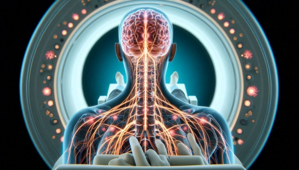
Paresthesia can be an unsettling experience. The tingling, prickling sensations or numbness in various parts of your body may leave you wondering about their origin. While many cases resolve on their own, understanding the underlying causes is essential for effective treatment. This is where advanced imaging techniques like MRI and CT scans come into play.
These tools are crucial in diagnosing paresthesia by providing detailed images of internal structures. They help healthcare providers pinpoint potential issues such as nerve compression or spinal abnormalities that could be contributing to your symptoms. By shedding light on the complexities behind paresthesia, MRI and CT scans pave the way for tailored treatments and improved patient outcomes.
Join us as we explore how these imaging studies work, when they’re necessary, and what you can expect during the process! Whether you’re experiencing unexplained sensations or just curious about medical diagnostics, this guide will equip you with valuable insights into the role of MRI and CT scans in identifying paresthesia causes.

Understanding MRI and CT Scans in Paresthesia Diagnosis
MRI and CT scans are vital tools in diagnosing paresthesia. These imaging techniques provide detailed insights into the body’s internal structures. Understanding how they work can demystify their role in identifying causes of abnormal sensations.
An MRI, or magnetic resonance imaging, uses powerful magnets and radio waves to create high-resolution images of soft tissues. This makes it ideal for visualizing nerves, muscles, and brain structures that may contribute to paresthesia symptoms.
On the other hand, a CT scan employs X-rays to generate cross-sectional images of bones and organs. It’s particularly useful for assessing spinal issues or fractures that might be compressing nearby nerves.
Both tests offer unique advantages depending on the clinical scenario. They help doctors not only confirm suspicions but also rule out various conditions that could lead to tingling or numbness in patients’ extremities. By leveraging these advanced technologies, healthcare professionals can develop more accurate treatment plans tailored to individual needs.
When Are Imaging Studies Necessary for Paresthesia?
Paresthesia, characterized by sensations like tingling or numbness, can arise from various underlying conditions. While many cases resolve on their own, some require further investigation through imaging studies. Understanding when these studies are necessary is key to effective diagnosis and treatment.
If paresthesia persists or worsens over time, it’s essential to consult a healthcare professional. Imaging may be indicated if there’s suspicion of nerve compression due to herniated discs, tumors, or other structural issues. This is particularly true for symptoms affecting the arms and legs.
Additionally, if paresthesia accompanies weakness, severe pain, or loss of bladder control, immediate imaging might be warranted. These signs could indicate serious neurological conditions that need prompt evaluation.
A thorough medical history and physical examination help determine the need for MRI or CT scans. Your doctor will assess whether your symptoms align with potential abnormalities visible through these advanced imaging techniques.
MRI Scans: Detailed Imaging of Soft Tissues and Nerves
MRI scans use powerful magnets and radio waves to create detailed images of the body’s soft tissues. This makes them particularly valuable in diagnosing conditions related to paresthesia, a sensation often described as tingling or numbness.
One significant advantage of MRI is its ability to visualize nerves and surrounding structures without exposing patients to radiation. This non-invasive technique provides clear images of brain, spinal cord, and peripheral nerve tissues.
Doctors can assess various factors contributing to paresthesia through these detailed scans. They look for signs of compression, inflammation, or lesions that may be affecting normal nerve function.
Additionally, MRIs can reveal issues such as herniated discs or tumors that could compress nearby nerves. By understanding how these elements interact within the body, healthcare providers can develop targeted treatment plans tailored to each patient’s unique needs.
CT Scans: Visualizing Bone Structures and Spinal Issues
CT scans, or computed tomography scans, are powerful diagnostic tools that excel at visualizing bone structures and detecting spinal issues. This imaging technique combines X-ray technology with advanced computer processing to create detailed cross-sectional images of the body.
For patients experiencing paresthesia—those tingling sensations often linked to nerve compression or injury—a CT scan can reveal critical information about the underlying skeletal framework. It allows healthcare providers to examine vertebrae for fractures, misalignments, or degenerative changes that may contribute to nerve problems.
CT scans are particularly effective in assessing conditions like herniated discs and spinal stenosis. These issues can compress adjacent nerves and lead to symptoms such as numbness or weakness in limbs. The clarity of a CT image aids doctors in pinpointing these abnormalities precisely.
The speed of a CT scan is another advantage; it typically takes only minutes to perform. Patients benefit from quick results that facilitate timely diagnosis and treatment plans tailored specifically for their condition.
Preparing for Your MRI or CT Scan: What You Need to Know
Preparing for an MRI or CT scan can seem daunting, but a little knowledge makes the process smoother. First, you should talk to your doctor about any medications you’re taking. Some may need to be paused before the procedure, especially if contrast dye is used.
Next, wear comfortable clothing without metal fasteners or zippers. These items can interfere with imaging quality and might require removal during the scan. Your facility may provide a gown for you to change into as well.
If you’re anxious about enclosed spaces, especially for an MRI, let your healthcare provider know beforehand. They might offer sedation options or allow someone to accompany you.
Follow instructions regarding food and drink intake prior to the appointment. For some scans like CTs using contrast material, fasting may be necessary for several hours leading up to your procedure for optimal results.
The Scanning Process: Step-by-Step Explanation
The scanning process for MRI and CT scans is straightforward but varies slightly between the two. First, you’ll arrive at the imaging center where a technician will guide you through the procedure. They’ll ask questions about your medical history and any concerns regarding allergies or previous reactions to contrast materials.
For an MRI, you’ll be asked to lie on a padded table that slides into a large magnet-shaped machine. It’s essential to remain still during the scan, which typically lasts 20 to 60 minutes. The machine produces loud noises, so earplugs or headphones are often provided.
In contrast, a CT scan involves lying on a narrow table that moves through a circular X-ray machine. This scan usually takes just a few minutes but may require you to hold your breath briefly while images are taken.
After both procedures, there’s no recovery time needed unless sedation was used in specific cases. You can resume normal activities shortly after leaving the facility.
Interpreting MRI Results: What Your Doctor Looks For
When your doctor reviews MRI results, they focus on specific aspects to pinpoint the cause of paresthesia. The primary goal is to identify any abnormalities in soft tissues and nerve structures. This includes looking for signs of inflammation, tumors, or cysts that could be compressing nerves.
Another critical area of assessment is the spinal column. Doctors examine the vertebrae and intervertebral discs for herniation or degeneration that might affect nerve pathways. Any structural changes can provide clues about pain and numbness sensations experienced by patients.
Your doctor will also consider surrounding blood vessels. Vascular issues may contribute to reduced blood flow, leading to symptoms like tingling or weakness in various body parts.
Qualitative data from the imaging helps determine if there’s a need for further investigation or immediate intervention. The interpretation process requires not only technical expertise but also an understanding of how these factors relate to your reported symptoms.
CT Scan Findings: Identifying Structural Abnormalities
CT scans are powerful diagnostic tools that provide detailed images of the body’s internal structures. When it comes to identifying causes of paresthesia, they play a crucial role in revealing structural abnormalities. These scans excel at visualizing bone and joint conditions that may lead to nerve compression.
Radiologists carefully examine CT scan results for signs of herniated discs, tumors, or fractures. A herniated disc can exert pressure on nearby nerves, causing sensations like tingling or numbness. Tumors—whether benign or malignant—can also impact nerve function by encroaching on surrounding tissues.
In addition to soft tissue evaluation, CT scans are adept at detecting spinal stenosis. This condition narrows the spinal canal and can result in various neurological symptoms. By pinpointing these issues, healthcare providers can create targeted treatment plans tailored to individual needs.
The clarity and precision offered by CT imaging allow for early intervention when necessary. Recognizing these structural problems is essential for addressing underlying causes contributing to paresthesia symptoms effectively.
Comparing MRI and CT Scans: Which is Best for Your Case?
When considering MRI and CT scans for diagnosing paresthesia, understanding their differences is crucial. MRIs excel at imaging soft tissues, providing detailed views of nerves and brain structures. This makes them highly effective for identifying conditions like multiple sclerosis or nerve compression.
On the other hand, CT scans shine in visualizing bone structures. They are often preferred when assessing fractures, tumors near bones, or spinal issues that could be causing symptoms. If your healthcare provider suspects a structural problem related to the skeletal system, a CT scan might be recommended.
The choice between MRI and CT also depends on factors such as patient comfort and time efficiency. MRIs take longer but provide more comprehensive information about soft tissue problems. Conversely, CT scans are quicker and can be performed rapidly in emergency situations.
Your doctor will consider your specific symptoms, medical history, and the suspected underlying cause to determine which imaging study is most appropriate for you.
Follow-Up Care: Understanding Your Imaging Results
Understanding your imaging results is a crucial step in addressing the underlying causes of paresthesia. After completing an MRI or CT scan, the next phase involves reviewing the findings with your healthcare provider. They will explain what the images reveal about your condition and how it relates to your symptoms.
It’s essential to discuss any abnormalities detected during the scans. Your doctor will clarify whether these issues could be contributing factors to your paresthesia. Depending on the results, further tests may be necessary for a comprehensive diagnosis.
If structural problems are identified—such as herniated discs or nerve compression—your care plan might include physical therapy, medication, or possibly surgery. Understanding these options empowers you to participate actively in decisions regarding your treatment path.
Don’t hesitate to ask questions during this follow-up appointment. Clear communication can help demystify complex medical jargon and provide peace of mind as you navigate this health concern. By grasping both your imaging results and their implications, you position yourself better towards effective management of paresthesia causes and improved quality of life.


